HPA axis and the autonomic nervous system
HPA axis and stress regulation
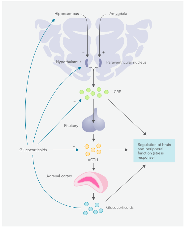
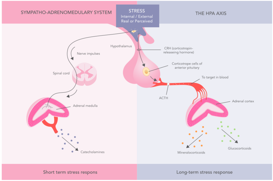
The HPA axis (hypothalamic-pituitary-adrenal) is the body's most important physiological feedback system for stress response. This is a neuroendocrine system, which means that hormones are secreted from certain parts of the system (e.g. hypothalamus and pituitary gland).
The operation and regulation of the HPA axis are influenced by the brain's nerve impulses and biochemical signals from different body parts. Particularly important brain structures affecting the stress response are the prefrontal cortex, the hippocampus, and the amygdala, anatomical structures, for example in the formation of memory and emotional reactions.
Their function is strongly linked to glucocorticoid secretion and stress-induced behavioral changes. The disorder between the higher parts of the brain and the HPA axis has been associated with various neuropsychiatric syndromes such as depression and post-traumatic stress disorder (PTSD). Damages in the prefrontal brain region and hippocampus, for example, can increase the susceptibility to strong and uncontrolled stress reactions.
When triggered by a stressor, the hypothalamus produces and secretes corticotropin-releasing hormone (CRH) and vasopressin, or anti-diuretic hormone (ADH). In particular, CRH (but also ADH) regulates the activity of the anterior pituitary by stimulating corticotropin (ACTH) secretion. Similarly, ACTH activates the adrenal cortex to secrete glucocorticoid hormones such as cortisol and also slightly to androgen dehydroepiandrosterone (DHEA) and mineralocorticoids such as aldosterone.
Like other endocrine systems, the HPA axis has a negative feedback system. Here, high blood cortisol levels reduce the hormone secretion of the pituitary and hypothalamus (CRH, ADH, and ACTH) and the hypothalamic stimulating effect through the glucocorticoid receptors (GR) in the cells (e.g. the brain). This way, your body avoids excessive or too low hormonal secretion, constantly striving for balance or homeostasis.
Glucocorticoids and cortisol in stress regulation
Glucocorticoids are part of the adrenal cortex corticosteroids. Their physiological effects are mediated through numerous glucocorticoid receptors (GR) in cells. They affect, inter alia, metabolism by promoting lipolysis (breaking down fat), increasing the release of amino acids and glucose into the bloodstream and preventing glucose from entering muscle and fat cells.
In addition, glucocorticoids affect the immune system, skeleton, cardiovascular system, reproduction, and cognitive function. Glucocorticoids, for example, strengthen myocardial contraction and reduce inflammation and the intensity of allergic reactions.
The most important glucocorticoid for the human body is cortisol, excreted approximately 15–30 mg daily from the middle part of the adrenal cortex (zona fasciculata). The body's stress reaction temporarily increases cortisol production, after which it usually returns to normal levels. In chronic stress conditions, cortisol production may remain permanently above normal levels. Like insulin resistance, this may cause glucocorticoid receptor resistance (GCR). Glucocorticoid receptor resistance predisposes the body to flu, and probably also to many other inflammatory diseases.
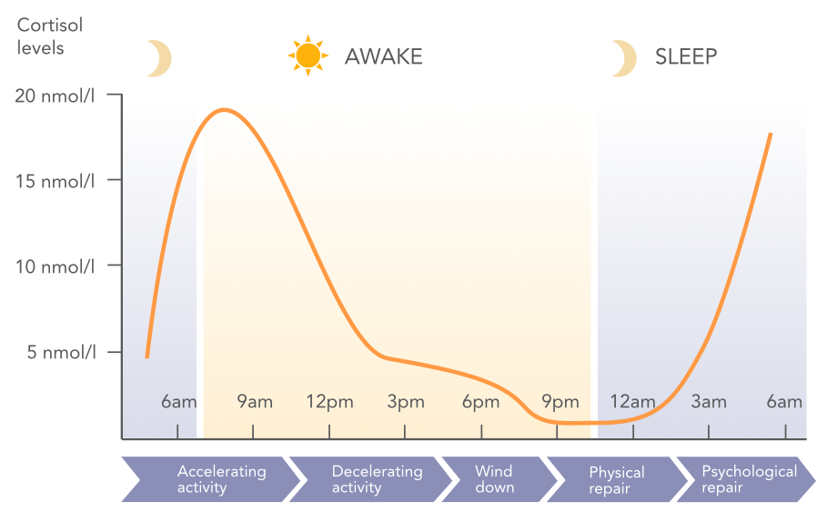
The excretion of cortisol from the adrenal glands is periodic (pulsed) - it follows both circadian (day-to-day) and ultradian (hours) rhythms. For example, in the morning, cortisol secretion is at its highest, helping to awaken and significantly increase alertness. Cortisol secretion gradually decreases towards the evening and is normally at its lowest around midnight. Studies have shown that this is one of the most stable circadian rhythms in compliance with the following phenomena in the body.
Pulse secretion is important for the subsequent physiological responses to occur normally. Animal experiments have shown that lack of pulsed cortisol secretion impairs hippocampus memory activity and often eliminates acute stress response. The disappearance of circadian rhythm has been associated with, for example, fatigue, sleep disorders, metabolic disorders and generally impaired quality of life.
The fight-or-flight response
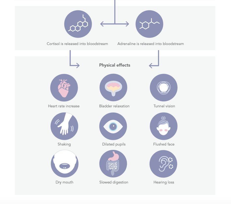
The fight-or-flight response is a physiological phenomenon triggered by various threats, situations, or thoughts. The significance of the term and phenomenon was first observed by Walter Bradford Cannon (1871-1945), Professor of Physics at Harvard University, in 1915. Cannon later also developed the term homeostasis, which refers to the attempt to restore body balance.
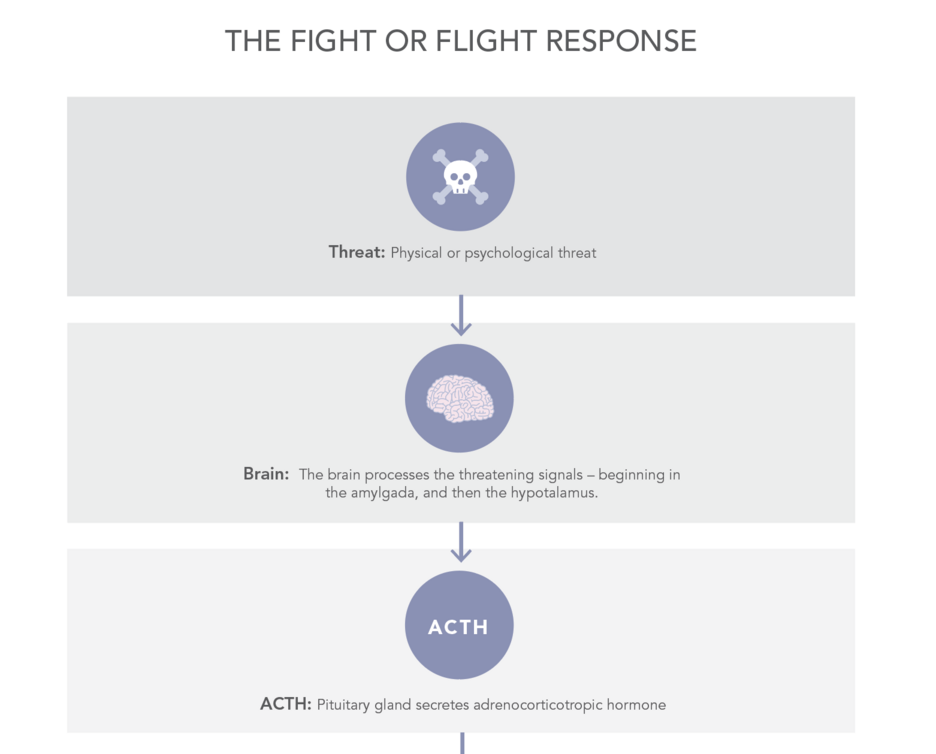
The "Fight or Flight" reaction is a mechanism, which is programmed into the nervous system of all mammals and acts as part of the normal defense system. As an example from nature take for example may b a zebra that freezes in place when seeing a lion. It has two alternatives: either to fight or escape. In humans, the fight-or-flight reaction is built into the "lizard brain" (according to the triune brain theory), ie. in the basal and deep parts of the brain, such as the amygdala, from which the signal progresses to the hypothalamus.
From there on, the body responds to the intensity of the stressor through the sympathetic nervous system and the HPA-axis: the higher the stimulus of the stressor, the stronger the body's response to it. According to a study published in 2016, in addition to the amygdala, there is a specific area called the Bed Nucleus of the Stria Terminalis (BNST) in the brain, whose role in the fight or flight reaction seems to be to mediate stress responses, especially in psychologically threatening situations.
If it is impossible to fight or escape, a paralysis reaction may occur. This biological protection mechanism protects people from the effects of a shocking situation. Some researchers have suggested that instead of a "fight or flight" reaction, this should be called a "fight, flight or freeze" reaction.
The fight or flight reaction is quite common in modern society's hectic and stressful lifestyle but is usually unnecessary. For example, the phenomenon may arise due to reading an angry e-mail or, for example, receiving a threatening call. Meditation has been found to produce a relaxation response that is the opposite of a fight-or-flight reaction.
The autonomic nervous system – the key to a healthy body
An autonomic nervous system is a non-volitional system regulating the body's vital functions. Autonomous (from the Greek words autos or self and nomos, or law) means independent and self-regulating. For the first time, an ancient Greek physician, psychologist, and experimental physiologist Claudio Galenos discovered an autonomic nervous system at the autopsy of pigs. The English physiologist John Newport Langley (1852–1925) developed the original term and definition of the autonomic nervous system in 1898 when investigating the autonomic system of internal organs.
The autonomic nervous system is structurally part of the central and peripheral nervous systems, but functionally it is a separate part of the nervous system. The autonomic nervous system controls and regulates the function of smooth muscles, glands, myocardium, and secreting epithelium (such as the stomach, respiratory organs, etc.). Its vital functions include blood circulation, digestion, hormone secretion, heat regulation, breathing, urine output, and reproduction. The central nervous system, in turn, consists of the brain and spinal cord.
The autonomic nervous system is controlled in the brain, especially by the limbic system which registers stress signals or stressors. Based on sensory, emotional, and thought information, the brain mediates activating or inhibitory signals through the hypothalamus to the parasympathetic or sympathetic nervous system. One of the most important tasks of the hypothalamus is to strike a balance between autonomic nervous system responses and hormone secretion.
The autonomic nervous system includes also the enteric nervous system. It is the nervous system of the digestive tract, also known as 'the second brain'. According to various estimates, the enteric nervous system (ENS) has 200 to 600 million neurons. Correspondingly, according to an estimate published in 2009, the most important part of the central nervous system (CNS), the brain, consists of about 86 billion neurons. The central and enteric nervous systems are in constant communication with each other.
Structure of the autonomic nervous system
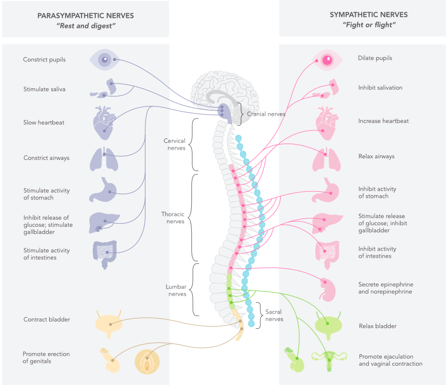
From the stress regulation point of view, the autonomic nervous system has two parts: a sympathetic nervous system and a parasympathetic nervous system. Alertness and stress reactions activate the sympathetic nervous system, whose active state is called sympathicotonus. On the other hand, the parasympathetic nervous system is responsible for the soothing state of the body (so-called parasympathicotonus), where the digestive system and the repair mechanisms of the various organs and cells are active.
However, the autonomous state of the body is rarely either sympathetic or parasympathetic but is often something between them (see picture below). Electrophysiological studies of the autonomic nervous system have shown that most organ nerves have sympathetic or parasympathetic nerve branches. Exceptions to this rule include iris, cardiac pacemaker cells, bladder, erectile tissue, salivary glands, oral mucosa, and intracranial vasculature, which are innervated with both sympathetic and parasympathetic nerve branches.
Vagus nerve - the conductor of the autonomic nervous system
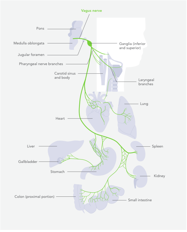
The Vagus nerve (the tenth cranial nerve) consists of two distinct nerve branches located on the right and left of the spinal cord. It is the most important and longest nerve in the parasympathetic nervous system. In practice, the vagus nerve regulates the function of all the internal organs, including the contraction of the muscles involved in speaking and eating.
The vagus nerve regulates, among other things, heart rate, bowel movements, sweating, speech output, eating and many other essential functions. Dysfunction of the vagus nerve may cause problems with digestion (dyspepsia, reflux, colitis, etc.), blood glucose control, urinary drainage, and immune function.
The vagus nerve also affects the functioning of the sympathetic nervous system through peripheral chemoreceptors. These senses especially the lack of blood oxygen in the peripheral parts and increase blood vessel contraction and increase blood pressure.